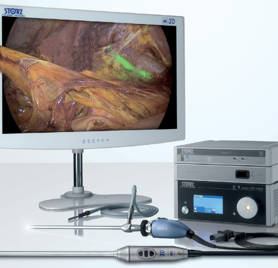
The use of NIR/ICG technology allows the visualization of various anatomical structures. Including:
• vascular system, hepatobiliary system and lymphatic system (with ICG administration).
• parathyroid glands (Without administration of ICG – endogenous autofluorescence)
• ureters (via an ICG-filled urethral catheter)
IMAGE1 S™ RUBINA components provide the user with new possibilities and a series of advantages, which aim to support them in their daily work.
When using Rubina components, you have several new modes for representing the NIR/ICG signal. These include NIR/ICG information overlaid on a standard white light image, as well as a mode that displays the pure infrared signal in a monochromatic color representation.
This is the Imaging of the future, with the Karl Storz guarantee.
Fazer login
Não tem conta? Registe-se


